Last Updated on July 1, 2025 by Luxe
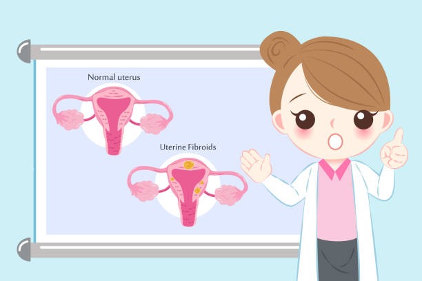
Uterine fibroids are non-cancerous growths that develop in or on the uterus and affect a significant number of women worldwide, especially those in their reproductive years. While some women may experience no symptoms at all, others face heavy menstrual bleeding, pelvic pain, and other complications that impact their quality of life. Because of the wide range of symptoms and the potential for confusion with other conditions, accurate diagnosis of uterine fibroids is essential.
If you suspect you might have uterine fibroids or have been experiencing symptoms like heavy periods, pelvic pressure, or frequent urination, it’s important to understand how uterine fibroids are diagnosed, and what tests and procedures your healthcare provider might use to confirm their presence.
In this blog, we’ll explore the common diagnostic tools and processes used to detect uterine fibroids, and explain what you can expect during each step.
Initial Medical History and Physical Exam
The diagnostic process often starts with your healthcare provider taking a detailed medical history. This discussion will include questions about:
- Your menstrual cycle (frequency, duration, and intensity),
- Symptoms such as pelvic pain or pressure,
- Urinary or bowel problems,
- Family history of fibroids or other uterine conditions,
- Fertility concerns.
Following this, a physical pelvic exam is usually performed. During this exam, the doctor will palpate (feel) your abdomen and pelvis to check for any irregularities, such as an enlarged or irregularly shaped uterus that might suggest fibroids.
Although a physical exam alone can’t confirm fibroids, it helps the doctor determine if further imaging tests are necessary.
Clinics like Advanced Gynaecology Melbourne, led by renowned gynaecologist Alex Ades, place great emphasis on thorough initial consultations to ensure no detail is overlooked.
Ultrasound Imaging
The most common and accessible test for diagnosing uterine fibroids is ultrasound imaging. Ultrasound uses sound waves to create a picture of the uterus and surrounding structures.
There are two types of ultrasound scans used:
- Transabdominal ultrasound: The probe is moved over your lower abdomen. This method provides a broader view of the uterus.
- Transvaginal ultrasound: A smaller probe is inserted into the vagina to get a closer and more detailed image of the uterus and endometrium.
Ultrasounds can detect the number, size, and location of fibroids, as well as differentiate fibroids from other uterine abnormalities such as cysts or polyps.
Advantages of ultrasound:
- Non-invasive and painless,
- Widely available and relatively affordable,
- No radiation exposure.
However, very small fibroids or those located deep in the uterine wall may sometimes be missed. At Advanced Gynaecology Melbourne, ultrasound assessments are a key part of the diagnostic process, often performed during the first visit to streamline your care.
Magnetic Resonance Imaging (MRI)
If ultrasound results are unclear, or if your healthcare provider needs more detailed information about your fibroids, an MRI scan may be recommended.
MRI uses powerful magnets and radio waves to produce detailed cross-sectional images of the uterus. It is particularly useful for:
- Differentiating fibroids from other uterine masses,
- Mapping the exact size and location of fibroids before surgery or treatment,
- Assessing the relationship of fibroids to the uterine cavity and other organs.
Though more expensive, MRI offers superior detail—making it a valuable tool in complex or multi-fibroid cases. Dr. Alex Ades frequently uses MRI at Advanced Gynaecology Melbourne for pre-surgical planning and personalised treatment strategies.
Hysterosonography (Sonohysterography)
Also known as saline infusion sonography, this specialized ultrasound procedure involves injecting sterile saline into the uterine cavity through a thin catheter inserted into the cervix. The saline expands the uterine cavity, allowing clearer visualization of the uterine lining.
This test is particularly helpful for:
- Detecting submucosal fibroids, which grow just beneath the uterine lining and can cause heavy bleeding or fertility issues,
- Evaluating the uterine cavity shape and size,
- Assessing the presence of polyps or adhesions.
Hysteroscopy
In this procedure, a thin, lighted telescope called a hysteroscope is inserted through the cervix into the uterus. It allows the doctor to look directly inside the uterine cavity.
Hysteroscopy is especially useful for:
- Diagnosing submucosal fibroids,
- Inspecting other abnormalities such as polyps or uterine septa,
- Sometimes allowing for simultaneous treatment like removing small fibroids or polyps.
Because hysteroscopy involves inserting an instrument into the uterus, it may require local or general anesthesia, depending on the case.
X-rays and Other Imaging
While less common for fibroid diagnosis, X-rays or computed tomography (CT) scans are sometimes used when other conditions need to be ruled out or when complex pelvic anatomy makes other imaging difficult.
These methods are generally not preferred due to lower detail compared to ultrasound or MRI and involve radiation exposure.
Blood Tests
Blood tests are not used to diagnose fibroids but can help evaluate symptoms and rule out other causes of heavy bleeding.
For example, your doctor may order:
- Complete blood count (CBC): To check for anemia caused by heavy menstrual bleeding,
Hormone tests: To evaluate hormonal imbalances that might affect fibroid growth, - Other labs: To rule out bleeding disorders or other conditions.
Biopsy
In rare cases, if a fibroid or uterine mass looks unusual or causes abnormal bleeding, your doctor might recommend an endometrial biopsy or even a more invasive tissue sampling to exclude cancerous changes.
When Should You See a Doctor?
If you experience any of the following symptoms, it’s important to consult a healthcare provider for evaluation:
- Heavy or prolonged menstrual bleeding,
- Pelvic pain or pressure,
- Frequent urination or difficulty emptying your bladder,
- Pain during intercourse,
- Unexplained anemia symptoms such as fatigue or weakness.
Early diagnosis and intervention can help avoid complications such as infertility or severe blood loss. Advanced Gynaecology Melbourne encourages patients to seek evaluation promptly to ensure timely and effective care.
Conclusion
Diagnosing uterine fibroids involves a combination of medical history, physical exams, and a range of imaging tests designed to give clear and detailed views of the uterus. The most common initial test is an ultrasound, but other advanced methods like MRI, hysterosonography, and hysteroscopy may be used to obtain more detailed information.
Understanding the tests and procedures involved can help ease anxiety and prepare you for what to expect during your diagnostic journey. If you suspect you have fibroids or are experiencing symptoms, make an appointment with your healthcare provider to discuss your concerns and receive an accurate diagnosis.